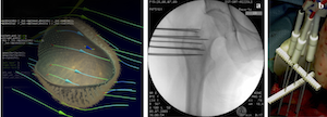
The use of electric field pulses to deliver therapeutic agents such as cytotoxic drugs and nucleic acids in cells and tissues has been successfully developed over the last decade. The method is currently used in clinics to treat skin cancers, a process called electrochemotherapy, and is also promising for vaccination and gene therapy. Its safe and efficient use for clinical purposes requires the knowledge of the mechanism underlying the DNA electrotransfer and expression phenomena. Despite the fact that the pioneering work on plasmid DNA electrotransfer in cells was initiated 30 years ago, many of the mechanisms underlying DNA electrotransfer remain to be elucidated. While efficient in vitro, the method faces a lack of efficiency in packed tissues such as tumors. Until now, the high majority of the studies have been performed on cells in 2D culture in Petri dishes or in suspension. However, these studies cannot get access to the tissue-specific architecture and organization present in 3D living tissues. In that context, 3D cell culture model are more relevant to in vivo cell organization since cell-cell contact are present as well as extracellular matrix. The aim of the presentation is to describe the relevance of using spheroids as a model to address and improve the electrotransfer processes.

|
|
|
Résumés par auteur > Golzio MurielHow 2D and 3D cell cultures can get access (or not) to the mechanisms of molecules delivery by pulsed electric field.
1 : Institut de pharmacologie et de biologie structurale
(IPBS)
-
Site web CNRS : UMR5089, Université Paul Sabatier [UPS] - Toulouse III, Université Paul Sabatier (UPS) - Toulouse III
205 Route de Narbonne 31077 TOULOUSE CEDEX 4 -
France
|
 PDF version
